

Hemangiosarcoma
Also known as: HSA, Canine hemangiosarcoma
In short
Hemangiosarcoma is a highly aggressive, malignant cancer of the blood vessels that commonly affects the spleen, heart, or skin in dogs and cats. Recognizing the signs of internal bleeding and understanding your diagnostic and treatment options can help you make informed decisions for your pet.
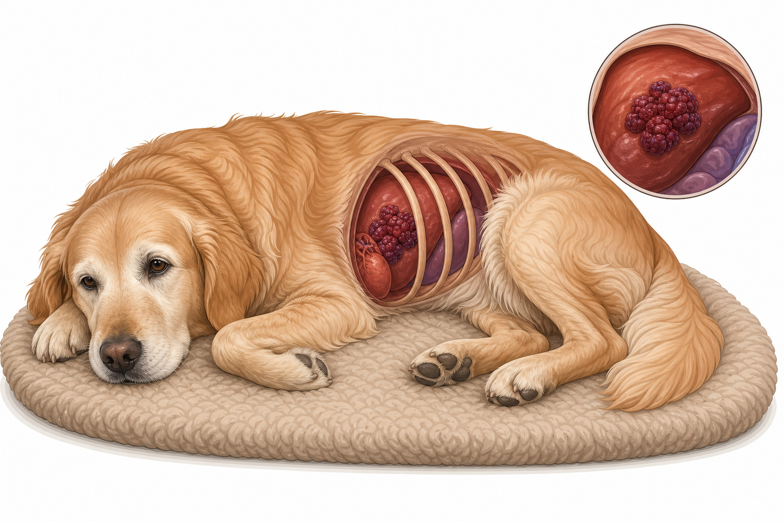
TL;DR. Hemangiosarcoma is a rapid, highly aggressive cancer of the blood vessels that primarily affects dogs, causing internal bleeding, sudden weakness, and requiring urgent veterinary intervention.
Hemangiosarcoma often presents with subtle signs of lethargy or weakness before a sudden crisis occurs.
What is it?
Hemangiosarcoma (HSA) is a highly aggressive, malignant cancer that originates in the endothelial cells, which are the cells that line the inside of blood vessels. Because blood vessels are present throughout the entire body, this cancer can theoretically develop anywhere. However, it has a strong preference for specific organs that are rich in blood supply.
When these cancerous cells multiply, they form abnormal, fragile blood vessels that do not hold blood properly. These malformed vessels are highly prone to leaking or rupturing entirely. This leads to sudden, life-threatening internal bleeding. The spleen is the most common site for this tumor to develop, but it frequently affects other vital organs as well.
According to a leading veterinary internal medicine reference:
"Approximately 50% of the tumors originate in the spleen, 25% in the right atrium, 13% in subcutaneous tissue, 5% in the liver, 5% in the liver-spleen–right atrium, and 1% to 2% simultaneously in other organs (i. e. , kidney, urinary bladder, bone, tongue, prostate)."
While hemangiosarcoma is most frequently diagnosed in dogs, cats can also develop this disease. In both species, the cancer is characterized by its ability to metastasize (spread) to other parts of the body very early in the course of the disease, often before any external signs of illness are visible.
Causes & risk factors
The exact underlying cause of internal hemangiosarcoma remains unknown. It is widely considered a multi-factorial disease where genetics play a primary role. There is no evidence that lifestyle, diet, or routine vaccinations influence the development of internal hemangiosarcoma.
However, for the cutaneous (skin) form of hemangiosarcoma, environmental factors play a clear role. Ultraviolet (UV) radiation from sun exposure is a major risk factor for dogs with light-colored skin and short coats. As noted in a standard veterinary dermatology guide:
"Solar damage may be involved in the development of tumors on the ventral glabrous skin in short-coated, lightly pigmented dogs (especially Whippets, Italian Greyhounds, Beagles, Great Danes, and English bulldogs), and on the head and ears"
Age is another significant risk factor. Hemangiosarcoma is principally diagnosed in middle-aged to older pets, with the risk increasing as they age.
Signs to watch for
The symptoms of hemangiosarcoma can vary dramatically depending on where the tumor is located and whether it has ruptured. Because the tumor grows silently inside the body, many pets show no signs of illness until the tumor ruptures and causes internal bleeding.
Cardinal Signs
- Anemia (Cardinal): This is the most consistent sign of hemangiosarcoma, caused by the ongoing loss of red blood cells into the abdomen or chest. You may notice your pet's gums look pale pink, grey, or completely white instead of a healthy bubblegum pink.
Common Signs
- Weakness: Your pet may suddenly seem lethargic, reluctant to rise, or unable to tolerate exercise.
- Spontaneous bleeding: This can manifest as nosebleeds, bruising on the skin, or prolonged bleeding from minor cuts.
- Hemoabdomen: Blood accumulation in the abdominal cavity, which often causes the belly to look distended or "pot-bellied."
- Acute collapse: A sudden loss of consciousness or inability to stand, often occurring when a tumor ruptures and causes a rapid drop in blood pressure.
- Thrombocytopenia: A severe drop in blood platelets, which are the cells responsible for helping the blood clot.
Occasional Signs
- Cardiac tamponade: Fluid or blood accumulation in the sac surrounding the heart (pericardium), which prevents the heart from beating effectively.
- Ventricular arrhythmias: Abnormal, irregular heart rhythms caused by tumors located in the heart muscle.
- Abdominal distention: A visibly swollen or heavy-feeling abdomen.
- Alopecia: Hair loss, which is occasionally seen in cutaneous forms of the disease.
- Ulceration: Open, bleeding, or non-healing sores on the skin, particularly in the cutaneous or subcutaneous forms.

Pale or white gums are a hallmark sign of severe anemia and internal bleeding, requiring immediate veterinary care.
One classic hallmark of internal hemangiosarcoma is a "waxing and waning" pattern of weakness. A pet may suddenly collapse or become very weak, only to recover and act normally a few hours or a day later. This happens when a small rupture occurs, causing minor internal bleeding that stops as the body's clotting mechanisms temporarily seal the leak. The body then reabsorbs the blood, and the pet feels better—until the next, often larger, rupture occurs.
How vets diagnose it
Diagnosing hemangiosarcoma requires a combination of blood tests, advanced imaging, and tissue analysis. Because the disease is highly aggressive, your vet will work quickly to determine the extent of the tumor and whether it has spread.
Your vet will start with a thorough physical exam and a complete blood count (CBC). A blood smear will be evaluated under a microscope to look for specific cellular changes. A leading veterinary internal medicine reference highlights these common blood findings:
"Most dogs with HSAs (83%) evaluated at the author's clinic are anemic; more than one half had RBC fragmentation and acanthocytosis. The pretreatment hemostasis profiles are normal in less than 20% of the dogs; most dogs (75%) have thrombocytopenia. Approximately one half of the hemostasis profiles meet three or more criteria for diagnosis of DIC."
These findings mean that the majority of affected dogs have damaged, fragmented red blood cells (schistocytes) and abnormally spiked red blood cells (acanthocytes) due to the physical trauma of squeezing through the tumor's malformed blood vessels. Furthermore, many dogs develop Disseminated Intravascular Coagulation (DIC), a catastrophic condition where the body's clotting factors are entirely consumed, leading to uncontrollable bleeding.
To locate the tumor and check for metastasis, your vet will utilize several imaging tools:
- Abdominal Ultrasound (AUS): This allows the vet to visualize the spleen, liver, and other abdominal organs to locate masses and detect free fluid (blood) in the abdomen.
- 3-View Chest Radiographs: X-rays of the chest from three different angles are essential to check for metastasis to the lungs.
- Echocardiography: An ultrasound of the heart is performed to look for tumors on the right atrium and to check for fluid in the heart sac.
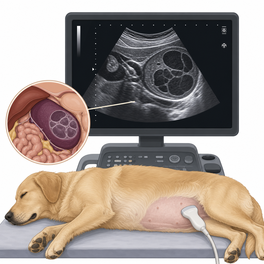
Abdominal ultrasound is a key diagnostic tool used to visualize masses on the spleen or liver.
While imaging and blood tests can strongly point to hemangiosarcoma, the gold standard for diagnosis is histopathology (biopsy). This involves sending the surgically removed tumor or a tissue sample to a veterinary pathologist for microscopic analysis. Fine-needle aspiration (FNA) and cytology (looking at cells collected via a needle) are often non-diagnostic because these tumors are highly vascular, meaning the sample is usually diluted with too much blood to find actual cancer cells.
Treatment options
Treatment for hemangiosarcoma is focused on stabilizing the pet, controlling bleeding, and slowing the spread of the cancer. Because this disease is highly aggressive, a multi-modal approach combining surgery and chemotherapy is typically recommended.
Surgery
For tumors located on the spleen or skin, surgical removal is the first line of defense. A splenectomy (complete removal of the spleen) is performed to eliminate the primary tumor and prevent further life-threatening internal bleeding. However, because micro-metastasis (the spread of microscopic cancer cells) has almost always occurred by the time of diagnosis, surgery alone is rarely curative for internal tumors.
Chemotherapy
To combat the rapid spread of the cancer, postoperative chemotherapy is highly recommended. The primary chemotherapy drugs used for hemangiosarcoma include:
- Doxorubicin: An anthracycline antineoplastic drug that is considered the cornerstone of most hemangiosarcoma chemotherapy protocols.
- Cyclophosphamide: An alkylating agent often used in combination with other drugs or as part of a low-dose, continuous (metronomic) chemotherapy protocol.
- Vincristine: A vinca alkaloid that may be incorporated into multi-drug chemotherapy regimens.
These treatments are designed to delay the progression of the disease while maintaining an excellent quality of life. Most dogs tolerate chemotherapy very well, with far fewer side effects than humans experience.
Supportive Care
Pets presenting in crisis often require immediate stabilization before any long-term treatment can be considered. This includes intravenous fluid therapy, blood transfusions (to treat severe anemia), and medications to manage pain and control ventricular arrhythmias.
Prognosis
Unfortunately, the long-term prognosis for pets with internal hemangiosarcoma is poor. The tumor is highly aggressive and spreads exceptionally fast.
Without treatment, survival times are very short, typically ranging from 20 to 60 days from the time of diagnosis, with death usually resulting from a catastrophic rupture and internal bleeding.
If surgery is performed to remove the primary tumor and is followed by a complete course of postoperative chemotherapy, survival times can be prolonged to approximately 140 to 202 days. While some pets do exceed these averages, owners should prepare themselves for a limited timeline and focus on maximizing their pet's quality of life.
For cats, hemangiosarcoma is less common, and long-term prognosis data in this species is limited. However, the disease behaves similarly aggressively in felines, though cutaneous forms that are completely excised early can sometimes carry a more favorable outlook.
Prevention
There is no known way to prevent internal hemangiosarcoma. Because the genetic factors are highly complex, there are currently no definitive genetic screening tests available for breeding dogs.
For the cutaneous (skin) form of the disease, prevention focuses on sun protection. You can protect high-risk dogs by:
- Limiting outdoor activity during peak sun hours (10:00 AM to 4:00 PM).
- Applying pet-safe, non-toxic sunscreen to lightly pigmented, hairless areas like the belly, nose, and ears.
- Using UV-blocking sun shirts for dogs that enjoy spending time outdoors.
For senior pets of highly predisposed breeds, routine screening—such as annual or bi-annual abdominal ultrasounds—can help detect splenic masses before they have a chance to rupture, allowing for controlled, elective surgery rather than an emergency crisis.
When to call your vet
Hemangiosarcoma can cause sudden, catastrophic emergencies. You should seek immediate emergency veterinary care if your pet exhibits any of the following signs:
- Sudden collapse or inability to stand
- Extremely pale or white gums
- Rapid, labored, or shallow breathing
- Sudden swelling or distention of the abdomen
- Extreme lethargy, weakness, or depression
- Unexplained bruising on the skin or bleeding from the nose or mouth
If your pet has already been diagnosed with hemangiosarcoma and is undergoing palliative care, these signs indicate a likely tumor rupture and require urgent veterinary assessment to ensure your pet's comfort.
For specific breeds
While any dog can develop hemangiosarcoma, certain breeds carry a significantly higher risk.
German Shepherd Dogs, Golden Retrievers, and Labrador Retrievers are highly predisposed to the abdominal form of the disease. As noted in a leading veterinary reference:
"Abdominal hemangiosarcoma is principally found in older dogs, especially German Shepherd Dogs and Golden Retrievers."
Other breeds with suspected predispositions to internal hemangiosarcoma include English Cocker Spaniels, English Setters, Afghan Hounds, Siberian Huskies, Irish Setters, and Great Danes.
Greyhounds have a unique presentation of this disease, frequently developing tumors within the muscles of their hind limbs. As described in veterinary literature:
"Greyhounds with intramuscular HSA typically present with a swollen and bruised rear limb; the tumor is frequently in the biceps femoris or quadriceps."
For short-coated, lightly pigmented breeds such as Whippets, Italian Greyhounds, Beagles, Great Danes, and English Bulldogs, the risk is highest for the cutaneous (skin) form of the disease due to solar damage on their sparsely haired bellies and ears.
Sources
- Small Animal Internal Medicine, 5th Edition, pages 529, 1220.
- Small Animal Dermatology: A Color Atlas and Therapeutic Guide, page 466.
My highlights & notes
Signs & symptoms
Breeds at higher risk
How it is diagnosed
- Histopathology / BiopsyGold standard
- 3-View Chest Radiographs
- Abdominal Ultrasound (AUS)
- Blood smear
- Echocardiography
- Fine-needle aspiration (FNA) / Cytology
- Hemostasis profile
Treatment approaches
Treatment must be prescribed by a licensed veterinarian based on your pet. Specific drug doses are intentionally not shown here.
Frequently asked questions
What is Hemangiosarcoma?
Hemangiosarcoma is a highly aggressive, malignant cancer of the blood vessels that commonly affects the spleen, heart, or skin in dogs and cats. Recognizing the signs of internal bleeding and understanding your diagnostic and treatment options can help you make informed decisions for your pet.
What are the symptoms of Hemangiosarcoma?
Anaemia (pale gums)、Acute collapse、Hemoabdomen、Spontaneous bleeding、Low platelets、Weakness (low energy, no strength)、Bloated belly、Hair loss
How is Hemangiosarcoma diagnosed?
Histopathology / Biopsy、3-View Chest Radiographs、Abdominal Ultrasound (AUS)、Blood smear、Echocardiography、Fine-needle aspiration (FNA) / Cytology
How is Hemangiosarcoma treated?
Treatment must be prescribed by a licensed veterinarian based on your pet. Specific drug doses are intentionally not shown here.
Sources
- Internal Medicine 5th · p. 1220
- Internal Medicine 5th · p. 1220
- 皮膚病 教科書點子書 Small-Animal-Dermatology-A-Color-Atlas-and-Therapeutic-Guide · p. 466
- Internal Medicine 5th · p. 529
- Internal Medicine 5th · p. 1220
This article is for general education and is not a substitute for professional veterinary advice. If your pet is unwell, please consult a veterinarian.
Worried about your pet?
Peqaboo’s AI helps you track symptoms, understand lab reports, and know when to see a vet.
Get the Peqaboo app