

Ventricular Septal Defect
Ventricular septal defect
Também conhecido como: VSD
Em resumo
A ventricular septal defect (VSD) is an uncommon congenital heart condition where an abnormal hole exists in the wall separating the heart's lower chambers, which can range from a harmless, self-closing minor defect to a serious risk for heart failure.
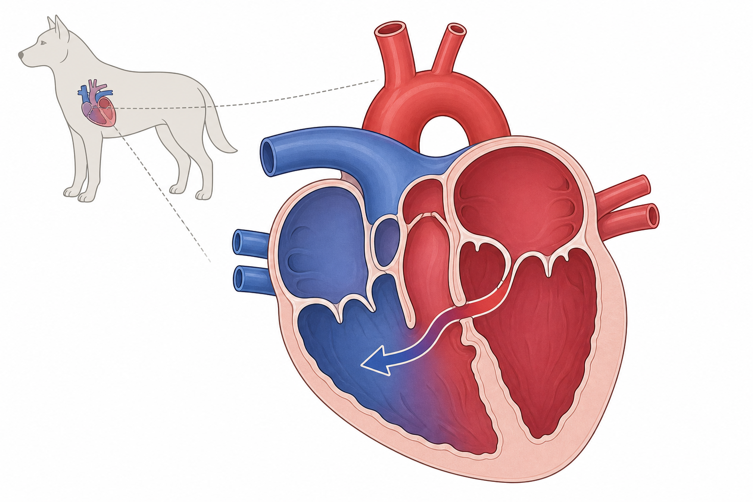
TL;DR. Um defeito do septo interventricular é um orifício congênito na parede entre os ventrículos; pode ser leve ou um risco sério de insuficiência cardíaca.
O VSD permite um shunt do ventrículo esquerdo de alta pressão para o direito.
O que é?
Malformação cardíaca congênita: orifício no septo interventricular. O sangue desvia da esquerda para a direita. O lado direito e os pulmões recebem volume excessivo; sobrecarga esquerda e insuficiência congestiva possíveis.
Causas e fatores de risco
Erro de desenvolvimento fetal com componente genético. Pouco frequente. Cães de risco: English Bulldog, English Springer Spaniel, Keeshond, West Highland White Terrier, Lakeland Terrier. Também em gatos.
Sinais a observar
- Sopro holossistólico (cardinal)
- Intolerância ao exercício (comum)
- Assintomático (comum) se pequeno/moderado
- Sopro de ejeção sistólica (ocasional)
- Insuficiência esquerda (ocasional): tosse, dispneia, letargia
- Sopro diastólico em decrescendo (raro)

A intolerância ao exercício é frequente em VSD grandes.
Diagnóstico
Ausculta. Confirmação: ecocardiografia [OURO], Doppler, radiografia torácica, ECG, cateterismo se necessário.
Tratamento
Nenhum fármaco fecha o orifício. Pequenos assintomáticos: vigilância. Se ICC/hipertensão pulmonar: diuréticos, vasodilatadores, suporte cardíaco. Fechamento espontâneo nos primeiros 2 anos possível.
"In some cases, the defect closes spontaneously within the first 2 years of life. Closure can result from myocardial hypertrophy around the VSD or a seal formed by the septal tricuspid leaflet or a prolapsed aortic leaflet."
Prognóstico
Pequeno/moderado: muitas vezes vida normal sob controle. Grande: reservado.
"A very large VSD causes the ventricles to function as a common chamber and induces RV dilation and hypertrophy. Pulmonary hypertension secondary to overcirculation is more likely to develop with large shunts."
Prevenção
Sem prevenção de estilo de vida. Não reproduzir afetados; triagem cardíaca dos pais em raças de risco.
Quando chamar o veterinário
Emergência: dispneia, respiração de boca aberta (sobretudo gato), tosse persistente, colapso, mucosas azuladas/pálidas, letargia extrema. Frequência respiratória em repouso muitas vezes <30/min.
Para raças específicas
Bulldog inglês, Springer inglês, Keeshond, Westie, Lakeland: ausculta cuidadosa no primeiro exame de filhote.
Fontes
- Internal Medicine, 5th Edition, pages 140, 141
- Clinical Echocardiography of the Dog and Cat, page 292
My highlights & notes
Sinais e sintomas
Raças de maior risco
Como é diagnosticado
- EchocardiographyPadrão-ouro
- Cardiac catheterization, oximetry, and angiocardiography
- Doppler echocardiography
- Electrocardiography
- Thoracic radiography
Perguntas frequentes
What is Ventricular Septal Defect?
A ventricular septal defect (VSD) is an uncommon congenital heart condition where an abnormal hole exists in the wall separating the heart's lower chambers, which can range from a harmless, self-closing minor defect to a serious risk for heart failure.
What are the symptoms of Ventricular Septal Defect?
Holosystolic murmur、No visible symptoms、Tiring easily during activity、Systolic ejection murmur、left-sided congestive heart failure、diastolic decrescendo murmur
How is Ventricular Septal Defect diagnosed?
Echocardiography、Cardiac catheterization, oximetry, and angiocardiography、Doppler echocardiography、Electrocardiography、Thoracic radiography
Fontes
- Clinical Echocardiography of the Dog and Cat · p. 292
- Internal Medicine 5th · p. 141
- Internal Medicine 5th · p. 140
- Internal Medicine 5th · p. 140
Este artigo é educação geral e não substitui orientação veterinária. Se o pet estiver mal, consulte um veterinário.
Preocupado com seu pet?
A IA da Peqaboo ajuda a registrar sintomas, entender exames e saber quando ir ao veterinário.
Baixar o app Peqaboo