

Ventricular Septal Defect
Ventricular septal defect
Juga dikenali sebagai: VSD
Ringkasnya
A ventricular septal defect (VSD) is an uncommon congenital heart condition where an abnormal hole exists in the wall separating the heart's lower chambers, which can range from a harmless, self-closing minor defect to a serious risk for heart failure.
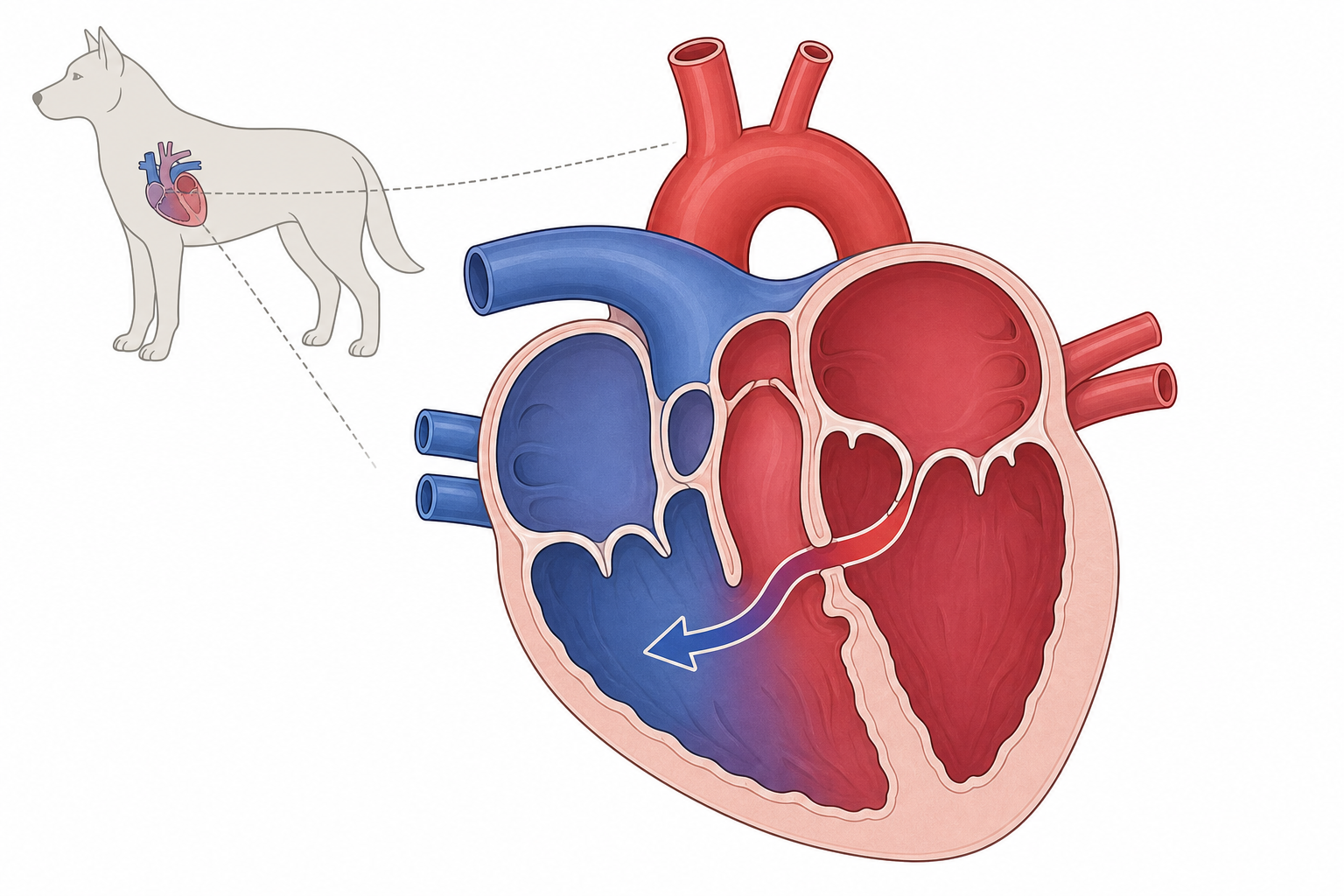
TL;DR. Kecacatan septum ventrikel ialah lubang kongenital pada dinding antara ventrikel; boleh ringan atau risiko serius kegagalan jantung.
VSD membolehkan shunt dari ventrikel kiri bertekanan tinggi ke kanan.
Apakah itu?
Malformasi jantung kongenital: lubang pada septum ventrikel. Darah mengalir kiri-ke-kanan. Sisi kanan dan paru menerima isipadu berlebihan; beban kiri dan kegagalan kongestif mungkin.
Punca dan faktor risiko
Ralat perkembangan janin dengan komponen genetik. Tidak lazim. Anjing berisiko: English Bulldog, English Springer Spaniel, Keeshond, West Highland White Terrier, Lakeland Terrier. Juga pada kucing.
Tanda
- Desiran holosistolik [kardinal]
- Intoleransi senaman [biasa]
- Asimptomatik [biasa] jika kecil/sederhana
- Desiran ejeksi sistolik [kadang-kadang]
- Kegagalan kiri [kadang-kadang]: batuk, dispnea, letargi
- Desiran diastolik decrescendo [jarang]

Intoleransi senaman biasa pada VSD besar.
Diagnosis
Auskultasi. Pengesahan: ekokardiografi [EMAS], Doppler, X-ray dada, ECG, kateterisasi jika perlu.
Rawatan
Tiada ubat menutup lubang. Kecil tanpa simptom: pantau. Jika CHF/hipertensi pulmonari: diuretik, vasodilator, sokongan jantung. Penutupan spontan dalam 2 tahun mungkin.
"In some cases, the defect closes spontaneously within the first 2 years of life. Closure can result from myocardial hypertrophy around the VSD or a seal formed by the septal tricuspid leaflet or a prolapsed aortic leaflet."
Prognosis
Kecil/sederhana: sering hidup normal di bawah kawalan. Besar: berwaspada.
"A very large VSD causes the ventricles to function as a common chamber and induces RV dilation and hypertrophy. Pulmonary hypertension secondary to overcirculation is more likely to develop with large shunts."
Pencegahan
Tiada pencegahan gaya hidup. Jangan biak yang terjejas; saringan jantung ibu bapa dalam baka berisiko.
Bila hubungi doktor haiwan
Kecemasan: sesak nafas, bernafas mulut terbuka (terutama kucing), batuk berterusan, rebah, membran biru/pucat, letargi melampau. Kadar nafas rehat sering <30/min.
Untuk baka tertentu
English Bulldog, English Springer Spaniel, Keeshond, Westie, Lakeland: auskultasi teliti pada pemeriksaan anak anjing pertama.
Sumber
- Internal Medicine, 5th Edition, pages 140, 141
- Clinical Echocardiography of the Dog and Cat, page 292
My highlights & notes
Tanda & simptom
Baka berisiko lebih tinggi
Cara didiagnosis
- EchocardiographyStandard emas
- Cardiac catheterization, oximetry, and angiocardiography
- Doppler echocardiography
- Electrocardiography
- Thoracic radiography
Soalan lazim
What is Ventricular Septal Defect?
A ventricular septal defect (VSD) is an uncommon congenital heart condition where an abnormal hole exists in the wall separating the heart's lower chambers, which can range from a harmless, self-closing minor defect to a serious risk for heart failure.
What are the symptoms of Ventricular Septal Defect?
Holosystolic murmur、No visible symptoms、Tiring easily during activity、Systolic ejection murmur、left-sided congestive heart failure、diastolic decrescendo murmur
How is Ventricular Septal Defect diagnosed?
Echocardiography、Cardiac catheterization, oximetry, and angiocardiography、Doppler echocardiography、Electrocardiography、Thoracic radiography
Sumber
- Clinical Echocardiography of the Dog and Cat · p. 292
- Internal Medicine 5th · p. 141
- Internal Medicine 5th · p. 140
- Internal Medicine 5th · p. 140
Artikel ini untuk pendidikan umum dan bukan pengganti nasihat doktor haiwan. Jika haiwan sakit, rujuk doktor haiwan.
Bimbang tentang haiwan kesayangan?
AI Peqaboo membantu jejak simptom, faham laporan makmal dan tahu bila jumpa doktor haiwan.
Muat turun app Peqaboo