

Ventricular Septal Defect
Ventricular septal defect
Ook bekend als: VSD
Kort samengevat
A ventricular septal defect (VSD) is an uncommon congenital heart condition where an abnormal hole exists in the wall separating the heart's lower chambers, which can range from a harmless, self-closing minor defect to a serious risk for heart failure.
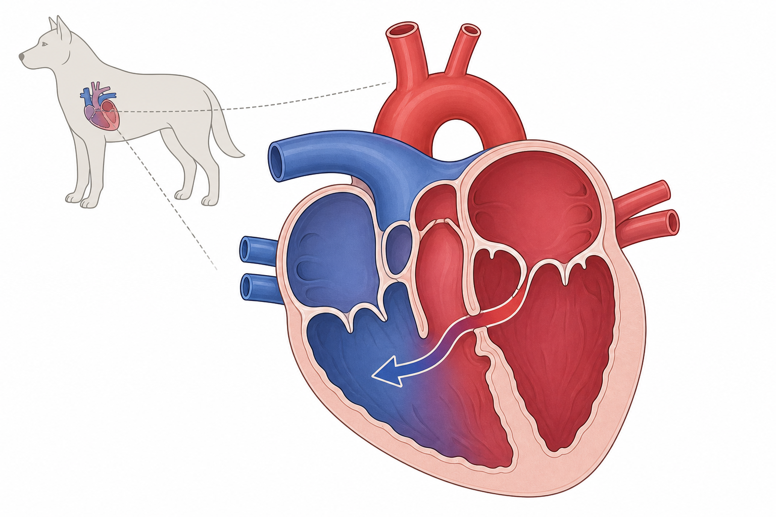
TL;DR. Een ventrikelseptumdefect is een aangeboren opening in de wand tussen de kamers; het kan mild zijn of een ernstig risico op hartfalen vormen.
Een VSD laat een shunt toe van de linker- (hoge druk) naar de rechterkamer.
Wat is het?
Aangeboren hartafwijking: gat in het ventrikelseptum. Bloed shunten links-naar-rechts. Rechterzijde en longen krijgen te veel volume; overbelasting links en congestief falen mogelijk.
Oorzaken en risicofactoren
Fout in foetale ontwikkeling, genetische component. Ongewoon. Risicohonden: English Bulldog, English Springer Spaniel, Keeshond, West Highland White Terrier, Lakeland Terrier. Ook bij katten.
Signalen
- Holosystolisch geruis (cardinaal)
- Inspanningsintolerantie (vaak)
- Asymptomatisch (vaak) bij klein/matig defect
- Systolisch uitdrijvingsgeruis (soms)
- Linkerhartfalen (soms): hoesten, dyspneu, lethargie
- Diastolisch decrescendo-geruis (zeldzaam)

Inspanningsintolerantie komt vaak voor bij grote VSD's.
Diagnose
Auscultatie. Bevestiging: echocardiografie [GOUD], Doppler, thoraxfoto, ECG, katheterisatie indien nodig.
Behandeling
Geen medicijn sluit het gat. Kleine asymptomatische: monitoring. Bij CHF/pulmonale hypertensie: diuretica, vasodilatoren, hartsteun. Spontane sluiting binnen 2 jaar mogelijk.
"In some cases, the defect closes spontaneously within the first 2 years of life. Closure can result from myocardial hypertrophy around the VSD or a seal formed by the septal tricuspid leaflet or a prolapsed aortic leaflet."
Prognose
Klein/matig: vaak normaal leven onder controle. Groot: gereserveerd.
"A very large VSD causes the ventricles to function as a common chamber and induces RV dilation and hypertrophy. Pulmonary hypertension secondary to overcirculation is more likely to develop with large shunts."
Preventie
Geen leefstijlpreventie. Getroffen dieren niet fokken; cardiale screening van ouders bij risicorassen.
Wanneer de dierenarts bellen
Spoed: ademnood, open-bekademhaling (vooral kat), aanhoudend hoesten, collaps, blauwe/bleke slijmvliezen, extreme lethargie. Ademfrequentie in rust vaak <30/min.
Voor specifieke rassen
English Bulldog, English Springer Spaniel, Keeshond, Westie, Lakeland: grondige auscultatie bij eerste puppycontrole.
Bronnen
- Internal Medicine, 5th Edition, pages 140, 141
- Clinical Echocardiography of the Dog and Cat, page 292
My highlights & notes
Tekenen & symptomen
Rassen met hoger risico
Hoe het wordt gediagnosticeerd
- EchocardiographyGouden standaard
- Cardiac catheterization, oximetry, and angiocardiography
- Doppler echocardiography
- Electrocardiography
- Thoracic radiography
Veelgestelde vragen
What is Ventricular Septal Defect?
A ventricular septal defect (VSD) is an uncommon congenital heart condition where an abnormal hole exists in the wall separating the heart's lower chambers, which can range from a harmless, self-closing minor defect to a serious risk for heart failure.
What are the symptoms of Ventricular Septal Defect?
Holosystolic murmur、No visible symptoms、Tiring easily during activity、Systolic ejection murmur、left-sided congestive heart failure、diastolic decrescendo murmur
How is Ventricular Septal Defect diagnosed?
Echocardiography、Cardiac catheterization, oximetry, and angiocardiography、Doppler echocardiography、Electrocardiography、Thoracic radiography
Bronnen
- Clinical Echocardiography of the Dog and Cat · p. 292
- Internal Medicine 5th · p. 141
- Internal Medicine 5th · p. 140
- Internal Medicine 5th · p. 140
Dit artikel is algemene educatie en geen vervanging voor professioneel veterinair advies. Bij ziekte: raadpleeg een dierenarts.
Zorgen over je huisdier?
Peqaboo’s AI helpt symptomen te volgen, labuitslagen te begrijpen en te weten wanneer je naar de dierenarts moet.
Download de Peqaboo-app