Pyoderma
Canine pyoderma
Also known as: Superficial pyoderma, Deep pyoderma, Bacterial skin infection, Superficial bacterial folliculitis, Chin pyoderma, Mucocutaneous pyoderma, Nasal pyoderma, Bacterial pododermatitis, Skin fold dermatitis
In short
Pyoderma is a common bacterial skin infection in dogs, ranging from superficial surface irritations to painful, deep tissue infections. Because it is almost always triggered by an underlying condition like allergies or hormonal imbalances, successful treatment requires both targeted antibiotics and addressing the root cause.
TL;DR. Pyoderma is a common bacterial skin infection in dogs that is almost always triggered by an underlying health issue, requiring targeted antibiotics and root-cause management for lasting relief.
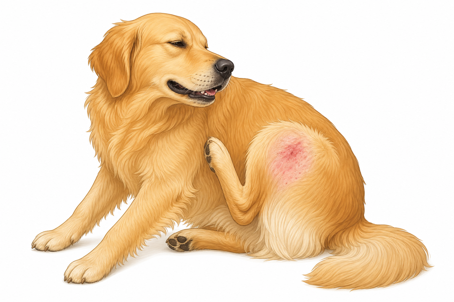
Pyoderma often starts with localized redness and itching, prompting dogs to scratch and further damage the skin barrier.
What is it?
Pyoderma is the medical term for a bacterial infection of the skin. Derived from the Greek words for "pus" (pyo) and "skin" (derma), it is one of the most frequent reasons dog owners seek veterinary care. While the word implies the presence of pus, canine pyoderma can manifest in many different ways, from simple dry flakes and hair loss to deep, oozing sores.
To understand pyoderma, it helps to look at how a dog's skin is structured. The canine skin barrier is a complex defense system made of skin cells, oils, and a natural population of microscopic organisms, including bacteria and yeast. Under normal conditions, these microbes live in harmony without causing harm. However, if the skin barrier is damaged, or if the dog's immune system is compromised, these normal bacteria—most commonly Staphylococcus pseudintermedius—multiply rapidly and invade the tissue, leading to infection.
Veterinarians classify pyoderma into two primary categories based on how deeply the infection penetrates the skin layers:
- Superficial Pyoderma: This form is confined to the upper layer of the skin (the epidermis) and the hair follicles. It is highly common and, while uncomfortable and itchy, is relatively straightforward to treat.
- Deep Pyoderma: This is a much more severe infection that breaks through the hair follicles to invade the deeper layers of the skin, including the dermis and subcutaneous tissue. As a leading veterinary internal medicine reference explains:
"Deep pyoderma is a surface or follicular bacterial infection that breaks through hair follicles to produce furunculosis and cellulitis. Its development is often preceded by a history of chronic superficial skin disease..." [1]
Understanding the depth of the infection is critical, as deep pyoderma requires much more aggressive, long-term treatment to prevent permanent scarring and systemic illness.
Causes & risk factors
It is vital for dog owners to understand that pyoderma is almost never a primary disease. Healthy canine skin is remarkably resistant to bacterial invasion. Therefore, when a dog develops pyoderma, it is nearly always a secondary symptom of an underlying "trigger" that has disrupted the skin's natural defenses.
Common underlying causes and risk factors include:
- Allergies: Environmental allergies (atopy), food allergies, and flea allergy dermatitis are the most common culprits. Allergies cause intense itching, leading the dog to scratch, chew, and lick their skin. This self-trauma damages the physical skin barrier, allowing bacteria to enter. As noted in veterinary dermatology literature, even localized trauma from scratching can quickly expand into a bacterial infection:
"An early superficial lesion (after clipping) on the lumbar region of a dog with flea allergy dermatitis. The papular perimeter suggests an expanding bacterial folliculitis." [3]
- Endocrine (Hormonal) Diseases: Conditions such as hypothyroidism (underactive thyroid) or Cushing's disease (hyperadrenocorticism) alter the structure and immune function of the skin, making it highly susceptible to bacterial overgrowth.
- Anatomical Factors: Dogs with heavy skin folds, such as English Bulldogs, trap moisture, heat, and friction in these crevices. This creates an ideal breeding ground for bacteria, a condition known as skin fold dermatitis.
- External Parasites: Fleas, mites (such as Demodex or Sarcoptes), and lice damage the skin directly and cause intense itching.
- Physical Trauma or Foreign Bodies: Foxtails, splinters, or pressure sores can introduce bacteria deep into the skin.
While any dog can develop pyoderma, certain breeds are highly predisposed due to their genetics, skin structure, or susceptibility to allergies. The English Bulldog and the German Shepherd Dog are two breeds with a well-documented predisposition to developing recurrent or deep forms of this condition.
Signs to watch for
Because pyoderma can affect different layers of the skin and occur anywhere on the body, its appearance varies widely.
Common Signs (Superficial Pyoderma)
- Pruritus (Itching): Dogs will scratch, bite, lick, or rub the affected areas constantly.
- Erythema (Redness): The skin will look pink, inflamed, or angry red.
- Papules and Pustules: Papules are small, raised red bumps. Pustules are classic "pimples" filled with yellow or white pus.
- Crusts and Scales: As pustules pop and dry, they form crusty scabs and flaky, peeling skin scales.
- Alopecia (Hair Loss): Hair often falls out in the infected areas. In short-coated breeds, this can look like a "moth-eaten" pattern.
- Lymphadenomegaly: Swollen lymph nodes, especially those near the site of a localized infection.
Occasional and Deep Pyoderma Signs
- Epidermal Collarettes: These are circular areas of peeling skin with a dark or hyperpigmented center, representing a ruptured pustule that has expanded outward.
- Moth-Eaten Patchy Alopecia: Particularly visible in short-haired dogs, where the coat looks uneven and patchy.
- Dull, Lusterless Hair Coat and Excessive Shedding: The overall quality of the coat degrades.
- Comedones: Blackheads, often seen in chronic cases or those associated with hormonal imbalances.
- Erosions and Ulcers: Raw, open sores where the top layers of skin have sloughed off.
- Pain and Swelling: Deep infections are highly painful to the touch, and the skin will feel thick and swollen.
- Draining Fistulous Tracts: Deep tunnels in the skin that ooze blood, pus, or clear fluid.
- Hemorrhagic Bullae: Large, blood-filled blisters.
- Cellulitis: Severe, widespread inflammation of the deep connective tissue.
- Systemic Signs (Fever, Depression, Anorexia, Lameness): If the infection enters the bloodstream or causes severe deep tissue destruction, your dog may become lethargic, lose their appetite, run a fever, or limp due to painful paws (pododermatitis).

Superficial pyoderma in short-coated dogs frequently causes a characteristic 'moth-eaten' pattern of hair loss and circular crusts.
How vets diagnose it
Diagnosing pyoderma itself is relatively straightforward, but identifying the exact type of bacteria and uncovering the underlying cause requires a systematic veterinary approach.
Your vet will start with a thorough physical examination and a detailed medical history. They will then perform basic dermatological tests to rule out other look-alike conditions, such as fungal infections (ringworm) or parasitic infestations (mites).
- Skin Scrapings: Your vet will gently scrape the surface of the skin with a scalpel blade to look for microscopic mites under the microscope.
- Impression Smears (Cytology): Your vet will press a glass microscope slide directly onto an active lesion, pustule, or moist skin area. They will then stain the slide and examine it under a microscope. This allows them to immediately identify whether bacteria (cocci or rods) or yeast are present, and confirm the presence of inflammatory cells (white blood cells).
- Gram Staining: This specialized staining technique helps categorize the bacteria on a microscopic level, guiding initial treatment decisions.
- Bacterial Culture and Sensitivity (The Gold Standard): If the infection is deep, recurrent, or has not responded to initial antibiotic therapy, a culture is absolutely essential. Your vet will sterilely collect a sample from a pustule, deep tissue, or draining tract. This sample is sent to a laboratory where the bacteria are grown and identified. Crucially, the lab tests various antibiotics against the bacteria to determine exactly which drugs will successfully kill them. As noted in diagnostic textbooks:
"Further characterization of bacterial organisms may be done with Gram staining, but definitive identification requires culture. Both aerobic and anaerobic culture should be considered for subcutaneous infections." [4]
- Anatomical Cultures: In cases of localized chronic infections, your vet may perform cultures from specific mucosal or junctional areas, such as the nose, lips, ears, axilla (armpits), and perianal areas, to identify bacterial reservoirs.
Treatment options
Successful treatment of pyoderma requires a multi-pronged approach: clearing the active bacterial infection, relieving the dog's immediate discomfort, and identifying and managing the underlying trigger.
First-Line Systemic Antibiotics
For superficial infections, your vet will often prescribe a first-line oral or injectable antibiotic. These are highly effective against standard skin bacteria and are chosen for their safety profile and ease of administration. Common first-line choices include:
- Cefpodoxime Proxetil: A convenient, once-daily third-generation cephalosporin antibiotic.
- Cefovecin: A long-acting injectable third-generation cephalosporin that provides continuous antibiotic coverage for up to 14 days, ideal for dogs that are difficult to medicate orally.
- Sulfadimethoxine/Ormetoprim: A potentiated sulfonamide antimicrobial that is highly effective for skin infections.
Second-Line Systemic Antibiotics
If a bacterial culture reveals that the infection is resistant to first-line drugs, or if the infection is deep and complex, your vet will step up to second-line therapies. These drugs are strictly reserved for resistant cases to prevent further bacterial resistance from developing.
- Orbifloxacin: A potent fluoroquinolone antibiotic that penetrates tissues deeply but must be used with caution and only when supported by culture results.
Topical Therapies
Topical therapy is a cornerstone of pyoderma management. In mild, superficial cases, topical therapy alone may be sufficient, avoiding the need for oral antibiotics entirely.
- Medicated Shampoos and Conditioners: Shampoos containing antibacterial agents (such as chlorhexidine or benzoyl peroxide) help physically wash away crusts, bacteria, and debris while directly killing microbes on the skin surface.
- Sprays, Mousses, and Ointments: These are useful for localized areas, such as the chin, paws, or skin folds.
Addressing the Root Cause
If the underlying allergy, parasite, or hormonal disease is not treated, the pyoderma will almost certainly return as soon as the antibiotic course is finished. Your vet will work with you to establish a long-term management plan for the primary disease, which may involve flea preventives, allergy medications, or hormone replacement therapy.
Prognosis
The prognosis for canine pyoderma is generally excellent, provided that the appropriate topical and systemic therapies are used for the correct duration, and the underlying predisposing factors are addressed. Superficial infections typically resolve within 3 to 4 weeks of treatment, though medications should always be continued for at least one week past the complete clinical resolution of symptoms.
Deep pyoderma requires a much longer course of treatment—often 6 to 12 weeks—and may result in localized scarring. If the underlying cause (such as severe atopic dermatitis or an unmanaged endocrine disease) cannot be fully resolved, the skin lesions may wax and wane, spontaneously resolve and recur, or persist indefinitely. In these chronic cases, lifelong topical maintenance therapy is often required to keep the bacterial population under control.
Prevention
While you cannot prevent the genetic factors that make some dogs prone to skin infections, you can take active steps to minimize the risk of pyoderma developing:
- Strict Parasite Control: Keep your dog on year-round, high-quality flea and tick preventives. Flea bites are a major trigger for the scratching cycle that leads to pyoderma.
- Manage Allergies Proactively: Work closely with your vet to manage environmental or food allergies. This may involve hypoallergenic diets, daily allergy medications, or immunotherapy.
- Regular Grooming and Bathing: For dogs prone to skin issues, regular bathing with a gentle, veterinary-recommended shampoo helps maintain a healthy skin barrier and washes away environmental allergens before they cause irritation.
- Skin Fold Care: If you own a breed with prominent skin folds, gently clean and dry these areas daily using veterinary-approved wipes to prevent moisture buildup.
- Prompt Attention to Minor Scratches: Clean minor cuts or scrapes promptly to prevent localized bacterial colonization.
When to call your vet
Pyoderma is not typically a sudden, life-threatening emergency, but it is highly uncomfortable and can escalate if left untreated. You should schedule a veterinary appointment if you notice your dog scratching excessively, losing hair, or developing red bumps and crusts.
You should seek immediate veterinary attention if your dog exhibits any of the following red-flag signs of deep, systemic infection:
- A high fever or extreme lethargy
- Loss of appetite (anorexia) or refusal to drink
- Severe, localized pain when the skin is touched
- Deep, open ulcers or widely spreading, purple-bruised skin (cellulitis)
- Pus or blood constantly draining from multiple holes in the skin
- Sudden lameness or inability to walk comfortably due to swollen, painful paws
For specific breeds
English Bulldogs
English Bulldogs are highly prone to skin fold dermatitis (intertrigo) due to their deep facial, tail, and body wrinkles. The lack of air circulation in these folds traps heat and moisture, leading to rapid bacterial overgrowth. Bulldogs are also prone to chin pyoderma (canine acne) and bacterial pododermatitis (painful bacterial infections between the toes). Daily hygiene of their skin folds and prompt treatment of any red bumps are essential for this breed.
German Shepherd Dogs
German Shepherds are uniquely predisposed to a severe, deep form of skin infection often referred to as German Shepherd Deep Pyoderma. This condition is believed to have an underlying hereditary or immunological component. It typically presents as painful, crusting, oozing ulcers and draining tracts on the outer thighs, groin, and trunk. Because of its deep nature, it requires aggressive, long-term antibiotic therapy, thorough diagnostic workups (including cultures), and careful management of any concurrent allergies.
Sources
- Small-Animal Dermatology: A Color Atlas and Therapeutic Guide, Page 48, 50, 54.
- Cowell and Tyler's Diagnostic Cytology and Hematology of the Dog and Cat, 5th Edition, Page 96.
My highlights & notes
Signs & symptoms
Breeds at higher risk
How it is diagnosed
- Bacterial culture and sensitivityGold standard
- Cultures from the nose, lips, ears, axilla, and perianal areas
- Gram staining
- Impression smears
- Scrapings
Treatment approaches
Treatment must be prescribed by a licensed veterinarian based on your pet. Specific drug doses are intentionally not shown here.
Frequently asked questions
What is Pyoderma?
Pyoderma is a common bacterial skin infection in dogs, ranging from superficial surface irritations to painful, deep tissue infections. Because it is almost always triggered by an underlying condition like allergies or hormonal imbalances, successful treatment requires both targeted antibiotics and addressing the root cause.
What are the symptoms of Pyoderma?
Hair loss、Skin redness、Papules、Itching and scratching、Pustules、Skin crusts、lymphadenomegaly、scales
How is Pyoderma diagnosed?
Bacterial culture and sensitivity、Cultures from the nose, lips, ears, axilla, and perianal areas、Gram staining、Impression smears、Scrapings
How is Pyoderma treated?
Treatment must be prescribed by a licensed veterinarian based on your pet. Specific drug doses are intentionally not shown here.
Sources
- 皮膚病 教科書點子書 Small-Animal-Dermatology-A-Color-Atlas-and-Therapeutic-Guide · p. 54
- 皮膚病 教科書點子書 Small-Animal-Dermatology-A-Color-Atlas-and-Therapeutic-Guide · p. 50
- 皮膚病 教科書點子書 Small-Animal-Dermatology-A-Color-Atlas-and-Therapeutic-Guide · p. 48
- Cowell and Tyler s Diagnostic Cytology and Hematology of the Dog and Cat, 5th Edition (VetBooks.ir) · p. 96
This article is for general education and is not a substitute for professional veterinary advice. If your pet is unwell, please consult a veterinarian.
Worried about your pet?
Peqaboo’s AI helps you track symptoms, understand lab reports, and know when to see a vet.
Get the Peqaboo app