

Cholangitis
Cholangitis
Also known as: Cholangiohepatitis, Neutrophilic cholangitis, Lymphocytic cholangitis, Suppurative cholangitis, Nonsuppurative cholangitis
In short
Cholangitis is a common and treatable inflammatory disease of the liver and bile ducts, primarily affecting cats. Learn about its bacterial and immune-mediated forms, key symptoms like jaundice, and how veterinarians diagnose and treat this complex condition.

TL;DR. Cholangitis is a common and treatable liver and bile duct disease in cats and dogs, requiring prompt veterinary care to manage inflammation or infection.

Early detection and veterinary care are key to managing cholangitis in cats.
What is it?
Cholangitis is a common inflammatory condition that targets your pet's biliary tract—the complex network of ducts that carry bile from the liver to the gallbladder and small intestine. While this disease can occasionally occur in dogs, it is a classic and highly significant disease in cats. When the inflammation spreads from the bile ducts into the surrounding liver tissue, veterinarians refer to the condition as cholangiohepatitis.
To understand cholangitis, it helps to look at how your pet's digestive system operates. The liver produces bile, a fluid essential for digesting fats. This bile travels through tiny intrahepatic bile ducts into the gallbladder, where it is stored. When your pet eats, the gallbladder contracts, sending bile through the common bile duct and into the small intestine. In cats, the common bile duct joins the pancreatic duct before entering the small intestine, sharing a single entry point. This close anatomical connection means that inflammation or bacterial overgrowth in the small intestine can easily migrate upward into both the pancreas and the biliary tract, often leading to concurrent inflammation of the intestines, pancreas, and liver—a syndrome vets call "triaditis."
Veterinarians classify cholangitis into two primary forms based on the types of inflammatory cells present:
- Neutrophilic (suppurative) cholangitis: This is typically an acute or chronic bacterial infection. Bacteria from the small intestine travel up the bile ducts, triggering a sudden, severe inflammatory response. White blood cells called neutrophils rush to the area to fight the infection.
- Lymphocytic (nonsuppurative) cholangitis: This is a chronic, slowly progressive form characterized by the infiltration of lymphocytes (a type of white blood cell associated with long-term immune responses) into the liver's portal areas. It is widely believed to be an immune-mediated condition, where the pet's own immune system mistakenly attacks the biliary tissues.
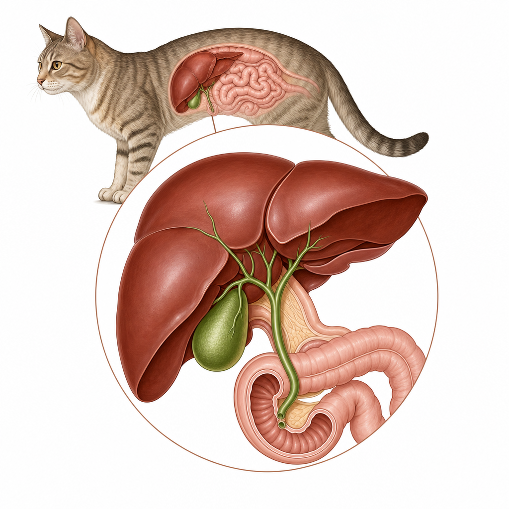
The close connection between the feline bile duct and pancreatic duct makes cats vulnerable to combined inflammation.
Causes & risk factors
The exact triggers for cholangitis depend heavily on the form of the disease:
- Bacterial Migration: Neutrophilic cholangitis is almost always caused by an ascending bacterial infection from the gastrointestinal tract. Common culprits include Escherichia coli, Enterococcus species, and Streptococcus species. Pets with pre-existing inflammatory bowel disease (IBD) or pancreatitis are at a much higher risk due to the shared anatomical pathways.
- Immune System Dysfunction: Lymphocytic cholangitis is thought to be an immune-mediated disease. The body's immune system becomes hyperactive, targeting the bile ducts and causing chronic, low-grade inflammation that can lead to scarring (fibrosis) over time.
- Breed Predispositions: Historically, Persian and Norwegian Forest Cats were thought to be predisposed to the lymphocytic form. However, modern veterinary research suggests that while these breeds may show historical associations, the disease is frequently diagnosed in older cats of any breed, with no clear, modern breed-wide predisposition.
As noted in a leading veterinary internal medicine reference:
"Cats with lymphocytic cholangitis were previously reported to be typically young to middle-aged, and Persians appeared to be overrepresented, but recent studies report it in older cats with no obvious breed predisposition (Callahan Clark et al, 2011; Warren et al, 2011)." [5]
Signs to watch for
The clinical signs of cholangitis can vary from subtle, chronic weight loss to sudden, life-threatening illness.
Common Signs
- Icterus (Jaundice): A yellow tinge to the gums, the whites of the eyes, or the skin, caused by a buildup of bilirubin in the bloodstream.
- Vomiting: Often frequent and accompanied by bile.
- Anorexia: A complete or partial loss of appetite.
- Dehydration: Indicated by dry gums or a loss of skin elasticity.
- Weight Loss: Especially noticeable in chronic lymphocytic cases.
- Lethargy: A general lack of energy or interest in normal activities.
Occasional Signs
- Abdominal Pain: Your pet may vocalize or tense their abdomen when picked up.
- Diarrhea: Often intermittent.
- Ascites: Fluid accumulation in the abdomen, making the belly look distended.
- Polyuria and Polydipsia: Increased urination and thirst.
- Fever: More common in acute neutrophilic infections.
- Hepatomegaly: An enlarged liver that your vet can feel during a physical exam.
Rare Signs
- Ptyalism: Excessive drooling, often secondary to severe nausea.
- Dullness: Mental depression or stupor.
- Seizures: Occurring in advanced stages if liver toxins build up and affect the brain (hepatic encephalopathy).

Icterus, or yellowing of the tissues, is a common sign of advanced biliary tract inflammation.
How vets diagnose it
Diagnosing cholangitis requires a systematic veterinary workup to differentiate between the neutrophilic and lymphocytic forms, as their treatments are vastly different.
- Serum Biochemistry: Blood tests typically reveal elevated liver enzymes (such as ALT, ALP, and GGT) and high levels of bilirubin. These elevations indicate active liver cell damage and compromised bile flow.
- Abdominal Ultrasonography: This non-invasive imaging tool allows your vet to evaluate the size and structure of the liver, check for gallbladder wall thickening, identify biliary sludge or gallstones, and rule out extrahepatic bile duct obstruction (a complete blockage of the main bile duct).
- Bile Cytology and Culture: Your vet may perform an ultrasound-guided cholecystocentesis to safely extract a small sample of bile directly from the gallbladder. This sample is analyzed under a microscope and cultured to identify any infecting bacteria, allowing for targeted antibiotic therapy.
- Liver Biopsy (The Gold Standard): A liver biopsy is the only definitive way to diagnose cholangitis and distinguish between the neutrophilic and lymphocytic forms. This can be performed via laparoscopy, laparotomy (surgery), or ultrasound-guided needle biopsy.
As described in a leading veterinary pathology reference:
"Lymphocytic cholangitis (LC) is a chronic form of disease that is characterized histologically by a mixed inflammatory infiltrate (typically small lymphocytes, or lymphocytes and plasma cells) within portal areas and is associated with varying degrees of fibrosis and bile duct hyperplasia." [2]
Treatment options
Treatment must be tailored to the specific type of cholangitis diagnosed. Misdiagnosing the type can lead to treatment failure; for example, giving immunosuppressive steroids to a pet with an active bacterial infection can be highly dangerous.
First-Line Therapies
- Antibiotics: Essential for treating neutrophilic cholangitis. Your vet will ideally select an antibiotic based on bile culture results. Aminopenicillins, such as amoxicillin or ampicillin, are common first-line choices. Metronidazole is often added to target anaerobic bacteria and provide mild anti-inflammatory benefits in the gut.
"Cats should be treated for 4 to 6 weeks with an appropriate antibiotic on the basis of the results of culture and sensitivity tests. Amoxicillin is a good initial choice..." [3]
- Bile Acids and Hepatoprotectants: Ursodiol (ursodeoxycholic acid) is widely prescribed to improve bile flow, dilute toxic bile acids, and reduce localized liver inflammation.
- Corticosteroids (Glucocorticoids): These are the cornerstone of treatment for lymphocytic cholangitis. Steroids suppress the abnormal immune response that drives chronic inflammation. However, they should only be started once a bacterial infection has been ruled out or successfully treated.
Second-Line and Supportive Therapies
- Nutritional Supplements: S-adenosylmethionine (SAMe) is a hepatoprotectant that supports liver cell regeneration and antioxidant production. Vitamin A supplementation may also be considered to support cellular health in chronic cases.
- Fluid Therapy: Intravenous or subcutaneous fluids are vital for correcting dehydration and maintaining electrolyte balance during acute flare-ups.
Prognosis
The prognosis for pets with cholangitis is generally good if the disease is diagnosed early and treated appropriately.
For acute neutrophilic cholangitis, many pets recover completely once the bacterial infection is eradicated. However, complications such as complete bile duct obstruction, pancreatitis, or systemic infection (sepsis) can significantly worsen the prognosis.
For lymphocytic cholangitis, the disease is chronic and slowly progressive. While a permanent cure is rare, many cats achieve long-term remission with consistent corticosteroid therapy and supportive care, enjoying an excellent quality of life for years. Regular veterinary follow-ups and blood monitoring are essential to manage chronic cases and adjust medication doses safely.
Prevention
Because the exact triggers of lymphocytic cholangitis remain unknown, and neutrophilic cholangitis typically stems from spontaneous bacterial migration from the gut, there are no guaranteed prevention strategies. Keeping your pet healthy, managing gastrointestinal diseases (like IBD) early, and scheduling regular veterinary checkups are the best defenses. There are currently no genetic screening tests available for predisposed breeds.
When to call your vet
If your pet is showing any signs of liver disease, prompt veterinary attention is vital.
Contact your veterinarian immediately or visit an emergency clinic if your pet exhibits:
- Yellowing of the gums, whites of the eyes, or skin (icterus/jaundice)
- Severe, persistent vomiting or inability to keep fluids down
- Extreme lethargy, collapse, or unresponsiveness
- Seizures or severe disorientation (signs of hepatic encephalopathy)
- Severe abdominal pain, often indicated by a hunched posture or crying out when touched
For specific breeds
If you own a Persian or Norwegian Forest Cat, pay close attention to subtle changes in their health. While modern studies show that any breed can develop cholangitis, historical data suggests these breeds may have an increased risk for the chronic, lymphocytic form. Regular wellness exams with routine blood screening can help catch early, silent liver inflammation before severe clinical symptoms develop.
Sources
- Small Animal Critical Care Medicine, 2nd Edition, page 679.
- Internal Medicine, 5th Edition, pages 577, 579, 580.
My highlights & notes
Signs & symptoms
Breeds at higher risk
How it is diagnosed
- Liver BiopsyGold standard
- Abdominal Ultrasonography
- Bile culture
- Bile cytology
- Serum Biochemistry
Treatment approaches
Treatment must be prescribed by a licensed veterinarian based on your pet. Specific drug doses are intentionally not shown here.
Frequently asked questions
What is Cholangitis?
Cholangitis is a common and treatable inflammatory disease of the liver and bile ducts, primarily affecting cats. Learn about its bacterial and immune-mediated forms, key symptoms like jaundice, and how veterinarians diagnose and treat this complex condition.
What are the symptoms of Cholangitis?
Loss of appetite、Dehydration、Yellowing of the skin and eyes (jaundice)、Lethargy、Vomiting (throwing up)、Weight loss (getting thinner)、Fluid in the belly (ascites)、Diarrhoea
How is Cholangitis diagnosed?
Liver Biopsy、Abdominal Ultrasonography、Bile culture、Bile cytology、Serum Biochemistry
How is Cholangitis treated?
Treatment must be prescribed by a licensed veterinarian based on your pet. Specific drug doses are intentionally not shown here.
Sources
- Internal Medicine 5th · p. 580
- Small Animal Critical Care Medicine, 2nd Edition (VetBooks.ir) · p. 679
- Internal Medicine 5th · p. 579
- Internal Medicine 5th · p. 577
- Internal Medicine 5th · p. 580
This article is for general education and is not a substitute for professional veterinary advice. If your pet is unwell, please consult a veterinarian.
Worried about your pet?
Peqaboo’s AI helps you track symptoms, understand lab reports, and know when to see a vet.
Get the Peqaboo app